2.75 recall that urine contains water, urea and salts
Urine contains:
- Salts
- Water
- Urea
Amount varies on the condition a person is operating
Salt + H2O affects the composition of tissue fluid
[Osmoregulation]
Urea - Excretion of metabolic waste
Monday, 7 November 2011
2.74 ADH
2.74 describe the role of ADH in regulating the water content of the blood
Anti diuretic hormone - ADH
Produced in the hypothalamus
- Flows to the bloodstream
- Target is the kidney
Effect of ADH
- Control and alter the quantity of water in the blood
- Ability to make the blood more or less concentrated
- Tissue fluid needs to be isotonic with the cytoplasm of the cell
ADH targets the collecting duct
- Allows more water to come out of the collecting duct
- Collecting duct ===selective reabsorption===> Blood
- ADH makes collecting duct walls more porous
- Water goes back into the blood
Consequence of ADH secretion
- Urine more concentrated
- Lower volume
ADH response to hot days, cold days, dehydration
2.73 Glucose reabsorption
2.73 understand that selective reabsorption of glucose occurs at the proximal convoluted tubule
Selective reabsorption of glucose
Reabsorption - Glomerular filtrate => Blood
End of the nephron: Urine
- Normally urine does not have glucose
- If there is glucose in the urine - diabetes
Proximal convoluted tubule (first)
- Glucose is removed
- Taken back into the blood

Selective reabsorption of glucose
Reabsorption - Glomerular filtrate => Blood
End of the nephron: Urine
- Normally urine does not have glucose
- If there is glucose in the urine - diabetes
Proximal convoluted tubule (first)
- Glucose is removed
- Taken back into the blood
2.72 Water reabsorption
2.72 understand that water is reabsorbed into the blood from the collecting duct
Bowman's Capsule - location of ultrafiltration
Dissolved contents of the blood are forced into glomerular filtrate
When filtration occurs - will filter out too much water therefore selective reabsorption occurs

Selective reabsorption of water occurs in the collecting duct:
Filtrate reaches the collecting duct
- Water is removed from the filtrate
- Returned back into the blood vessels
- Back into the bloodstream
- Selected and reabsorbed into the blood
Bowman's Capsule - location of ultrafiltration
Dissolved contents of the blood are forced into glomerular filtrate
When filtration occurs - will filter out too much water therefore selective reabsorption occurs
Selective reabsorption of water occurs in the collecting duct:
Filtrate reaches the collecting duct
- Water is removed from the filtrate
- Returned back into the blood vessels
- Back into the bloodstream
- Selected and reabsorbed into the blood
2.71 Ultrafiltration
2.71 describe ultrafiltration in the Bowman’s capsule and the composition of the glomerular filtrate
Nephron - Structure which carries out the filtration of blood
Produces:
- Filtered blood ("cleaned blood")
- Urine (the waste)
Urine - Water (H2O), salts, urea (N)
Beginning of the nephron structure begins at the Bowman's capsule
Where ultrafiltration begins
Filtration of blood:
1. Blood arriving in the kidney
Afferent arteriole
High pressure (come from an artery)
2. Blood leaving the kidney
Efferent arteriole
Narrow blood vessel (smaller than afferent arteriole)
3. Afferent ==> Efferent
Blood develops a high pressure
Blood pressure increases in glomerulus
4. High pressure forces the liquid in blood (plasma, which includes H2O, salts, amino acids, glucose, urea) into the inside of the Bowman's capsule (and consequently into the glomerula filtrate)
5. Glomerula filtrate
Plasma contains H2O, salts, amino acids, glucose, urea
Blood has been filtered by high pressure
Forces the liquid into the tube
 |
| Source: http://click4biology.info/c4b/11/11.3/glomerulus.gif |
2.70 Nephron Structure
2.70 describe the structure of a nephron, to include Bowman’s capsule and glomerulus, convoluted tubules, loop of Henlé and collecting duct
Nephron - functional unit of the kidney (filtration, controlling of composition of blood)
Aorta - taking blood into the kidney (renal artery)
Urine goes down into the ureters and into the bladder for excretion
Filtered blood goes back into the bloodstream through the renal vein (returns to the vena cava)
- Cortex
- Medulla
- Pelvic region - Space where the urine collects and drains down into the ureter
Kidney made up of millions of tubular structures (nephrons)
Tube starts on the edge of the medulla - dead end structure shown in diagram

Bowman's capsule - the dead end structure

Convoluted tubules - twisted sections
Dip in the nephron - Loop of Henlé
Bowman's Capsule - contains tight knot blood vessels [Glomerulus]
First twisted section - PCT (Proximal convoluted tubules)
Second twisted section - DCT (Distal convoluted tubules)
Nephron - functional unit of the kidney (filtration, controlling of composition of blood)
Aorta - taking blood into the kidney (renal artery)
Urine goes down into the ureters and into the bladder for excretion
Filtered blood goes back into the bloodstream through the renal vein (returns to the vena cava)
- Cortex
- Medulla
- Pelvic region - Space where the urine collects and drains down into the ureter
Kidney made up of millions of tubular structures (nephrons)
Tube starts on the edge of the medulla - dead end structure shown in diagram
Bowman's capsule - the dead end structure
Nephron - Tubular structure
Convoluted tubules - twisted sections
Dip in the nephron - Loop of Henlé
Bowman's Capsule - contains tight knot blood vessels [Glomerulus]
First twisted section - PCT (Proximal convoluted tubules)
Second twisted section - DCT (Distal convoluted tubules)
 |
| Source: http://www.beltina.org/pics/nephron.jpg |
Friday, 28 October 2011
2.69 Urinary system
describe the structure of the urinary system, including the kidneys, ureters, bladder and urethra
1. Right and left kidney
Each with its own separate blood supply
Excretion, filtration, osmoregulation
2. Ureter
Carries urine from the kidney to the bladder
Each kidney has a tube that leads into the common bladder
3. Bladder
Stores urine
4. Urethra
Where urine is carried out of the body
Travels down through either the vagina or the penis
 |
Source: www.biochem.co, www.medicalartlibrary.com |
1. Right and left kidney
Each with its own separate blood supply
Excretion, filtration, osmoregulation
2. Ureter
Carries urine from the kidney to the bladder
Each kidney has a tube that leads into the common bladder
3. Bladder
Stores urine
4. Urethra
Where urine is carried out of the body
Travels down through either the vagina or the penis
2.68b Osmoregulation
understand how the kidney carries out its roles of excretion and of osmoregulation
The kidney can control the amount of water and salts in the bloodstream, keeping the blood and tissue fluid isotonic and therefore maintaining the function of the cells
Osmoregulation
Osmo - osmosis (less conc. ==> more conc.)
Regulation - control

[1]
The blood circulating into the tissue could be very concentrated or dilute, causing the tissue fluid to be hypertonic or hypotonic - both of these conditions could remove or add too much water to the cells, causing the cells to lose its function
[2]
The kidneys keep the tissue fluid isotonic - controlling the composition of the blood
(Blood forms the tissue fluid)
Excess water and salts removed and excreted down into the ureter
The kidney can control the amount of water and salts in the bloodstream, keeping the blood and tissue fluid isotonic and therefore maintaining the function of the cells
Osmoregulation
Osmo - osmosis (less conc. ==> more conc.)
Regulation - control
[1]
The blood circulating into the tissue could be very concentrated or dilute, causing the tissue fluid to be hypertonic or hypotonic - both of these conditions could remove or add too much water to the cells, causing the cells to lose its function
[2]
The kidneys keep the tissue fluid isotonic - controlling the composition of the blood
(Blood forms the tissue fluid)
Excess water and salts removed and excreted down into the ureter
2.68a Excretion
understand how the kidney carries out its roles of excretion and of osmoregulation
Both the liver and kidneys remove toxic amino acids and urea from the body by breaking the amino acids down into urea (in the liver) and converting the urea into urine (in the kidneys), finally excreting it out of the body through the bladder in the form of urine
Urea, contains nitrogen
Nitrogen toxic to body - unable to store
Amino acids - Normally used for growth
Excess amino acids must be excreted (liver and kidneys)
1. Liver
- Blood circulates to the liver
- Amino acids in blood broken down
- Converted into urea
- Returns to the bloodstream
2. Kidneys
- Blood circulates to both kidneys
- Kidneys filter urea from blood
- Urea added to water to form urine
- Urine flows down into the bladder via ureters
- Filtered blood returns to the bloodstream in veins without toxic amino acids and urea
2.67b Human organs of Excretion
recall that the lungs, kidneys and skin are organs of excretion
1. Lungs
- Carbon dioxide (waste from respiration)
2. Kidneys
- Excess H2O
- Urea (nitrogen waste from amino acids, because of inability to store amino acids)
- Salts
3. Skin
- Sweat (H2O and salts)
- to a lesser extent, urea

1. Lungs
- Carbon dioxide (waste from respiration)
2. Kidneys
- Excess H2O
- Urea (nitrogen waste from amino acids, because of inability to store amino acids)
- Salts
3. Skin
- Sweat (H2O and salts)
- to a lesser extent, urea
2.67a Excretion in plants
recall the origin of carbon dioxide and oxygen and waste products of metabolism and their loss from the stomata of a leaf
Plants excrete oxygen (O2) and carbon dioxide (CO2) depending on whether they are carrying out photosynthesis or respiration
1. Photosynthesis
- Leaf absorbing light energy, producing oxygen
- CO2 + H2O -> C6H12O6 + O2 (Excretion - the release of metabolic waste)
- Waste excreted through the stomata
2. Respiration
- C6H12O6 + O2 -[Enzyme reactions]-> ATP + CO2 + H2O
Aerobic respiration
CO2 is metabolic waste - excretion
Monday, 10 October 2011
3.30 Mutation
Rare, random change in genetic material that can be inherited
Mutation changes the base sequence of the genes (e.g. ACT to AAT)
Allele - form of gene
Alleles exist because of mutation
i.e. A and a (dominant and recessive genes)
Mutation creates a new version of alleles
Creates new protein
Entirely different effect on phenotype
Mutation changes the base sequence of the genes (e.g. ACT to AAT)
Allele - form of gene
Alleles exist because of mutation
i.e. A and a (dominant and recessive genes)
Mutation creates a new version of alleles
Creates new protein
Entirely different effect on phenotype
3.29 Species Variation
Variation within a species can be
- Genetic
- Environmental
- Both
Variation -> differences in phenotype (determined by Variation = Genotype + Environment)
[Continuous variation]
 1. Variation entirely dependent on genotype.
e.g. blood group (A, AB, B, O...)
 2. Variation modified by the environment
e.g. tallness (inherited with genotype but diet may influence it)
3. Environmental variation
Entirely dependent on environment
Cannot be inherited genetically
e.g. home language
- Genetic
- Environmental
- Both
Variation -> differences in phenotype (determined by Variation = Genotype + Environment)
[Continuous variation]

e.g. blood group (A, AB, B, O...)

e.g. tallness (inherited with genotype but diet may influence it)
3. Environmental variation
Entirely dependent on environment
Cannot be inherited genetically
e.g. home language
3.31 Evolution
describe the process of evolution by means of natural selection
Evolution - Change in the form of organisms / Change in the frequency of alleles
Natural selection - Mechanism of evolution (Proposed by Charles Darwin)
Change in allele frequency by means of natural selection
-> e.g. Skin / lung infections by Staphylococcus aureus
Original form is susceptible to being killed by metheciline (antibiotic)
Random mutation - gives it ability to break down metheciline (resistant)
2 forms - (1st definition of evolution)
2nd form increases because the 1st form dies out

Natural selection:
Evolution - Change in the form of organisms / Change in the frequency of alleles
Natural selection - Mechanism of evolution (Proposed by Charles Darwin)
Change in allele frequency by means of natural selection
-> e.g. Skin / lung infections by Staphylococcus aureus
Original form is susceptible to being killed by metheciline (antibiotic)
Random mutation - gives it ability to break down metheciline (resistant)
2 forms - (1st definition of evolution)
2nd form increases because the 1st form dies out
Natural selection:
- Random mutation - Produces MRSA form
- Non-random selection - Antibiotic selecting MRSA to be killed
3.33 Antibiotic resistance
Understand how resistance to antibiotics can increase in bacterial populations
Staphylococcus aureus
- Skin infections
- Lung infections
Can be treated with metheciline
Antibiotic - Chemical can kill staph.
Type of staph. that can be killed with metheciline is called the susceptible form
MSSA - metheciline susceptible staph. aur.
----
Random mutation to the genotype of the staph.
Bacteria does not die when given methe.
Resistant
MRSA - metheciline resistant staph. aur.
----
Effects of use of antibiotics
MRSA increasing survival rate
Becomes more common
Resistance becoming an increasing problem within hospitals
3.34 Causes of mutation
Mutation - change of base sequence
1. Radiation
Ionising radiation (e.g. gamma rays, X-rays, ultraviolet UV-B)
UV-B causes mutation causing skin cancer
2. Chemicals
Tars, tobacco
Mutagens - Chemicals which cause mutations
Carcinogens - Mutagens which also cause cancer
1. Radiation
Ionising radiation (e.g. gamma rays, X-rays, ultraviolet UV-B)
UV-B causes mutation causing skin cancer
2. Chemicals
Tars, tobacco
Mutagens - Chemicals which cause mutations
Carcinogens - Mutagens which also cause cancer
3.32 Types of Mutation
Understand that many mutations are harmful but some are neutral and few are beneficial
Gene ==> New alleles [mutation]
Alleles are responsible for phenotype
New phenotype could be:
- Beneficial e.g. enzyme
- Neutral (neutrality may not last forever - +environmental changes may affect it and turn it beneficial or neutral)
- Harmful e.g. non functional enzyme
Gene ==> New alleles [mutation]
Alleles are responsible for phenotype
New phenotype could be:
- Beneficial e.g. enzyme
- Neutral (neutrality may not last forever - +environmental changes may affect it and turn it beneficial or neutral)
- Harmful e.g. non functional enzyme
Tuesday, 4 October 2011


3.20 Family Pedigrees

Finding out if the condition is caused by a dominant or recessive allele
Write out genotypes and look for evidence in the diagram:

3.18c Codominance
3rd (distinct) phenotype appears: Blue x White -> Orange
Codominance -> B = W both contribute to phenotype

Codominance -> B = W both contribute to phenotype
Monday, 26 September 2011


Sunday, 25 September 2011


Friday, 16 September 2011
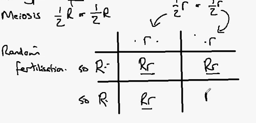
3.2 Fertilisation
Understand that fertilisation involves the fusion of a male and female gamete to produce a zygote that undergoes cell division and develops into an embryo

Adult male + Adult female
Diploid cells (2n) have complete set of chromosomes
In humans 2n = 46
Diploid cells divide (with meiosis) to create gametes with haploid set (n - ½ a set; 23 in humans)
Gametes in male - sperm
Gametes in female - egg

Fertilisation
Sexual reproduction
Cells are brought together - joined/fused together
Forms one cell (Chromosomes: n + n = 2n)
New cell has 46 chromosomes - Zygote
Combination of male and female chromosomes

Zygote goes through mitosis - Cells divide to give two cells
Both contain 46 chromosomes
All cells have 2n / diploid number of chromosomes
Sufficient cells -> creates embryo
Adult male + Adult female
Diploid cells (2n) have complete set of chromosomes
In humans 2n = 46
Diploid cells divide (with meiosis) to create gametes with haploid set (n - ½ a set; 23 in humans)
Gametes in male - sperm
Gametes in female - egg
Fertilisation
Sexual reproduction
Cells are brought together - joined/fused together
Forms one cell (Chromosomes: n + n = 2n)
New cell has 46 chromosomes - Zygote
Combination of male and female chromosomes
Zygote goes through mitosis - Cells divide to give two cells
Both contain 46 chromosomes
All cells have 2n / diploid number of chromosomes
Sufficient cells -> creates embryo
3.9b Female Reproductive System

Uterus generally small (size of orange) before pregnancy
1. Ovary - Meiosis - produces gametes (eggs)
2. Oviducts - Carry eggs to uterus
- Location where fertilisation takes place (in green)
3. Wall of uterus (muscle) - stretches during pregnancy and contracts during childbirth
4. Lining of uterus - accepts and develops egg -> embryo -> child (placenta)
5. Cervix - entrance to uterus for sperm
6. Uterus space - place where embryo develops
7. Vagina - Collects sperm cells
3.9a Male Reproductive System
Recall the structure and function of the male and female reproductive systems

1. Bladder - Stores urine
2. Testis - Meiosis (produces gametes - sperm cells)
3. Epididymis - Stores sperm cells
4. Vas deferens - Carries sperm cells to penis (tube)
5. Prostate - adds 20/30% of volume of semen, contains sugars, alkali (neutralise acidic secretions within vagina)
6. Seminal Vesicles - 70% of semen, contains "
Sperm cells are combined with prostate and seminal vesicle secretions to create semen
7. Urethra - Takes semen down penis, also exit for urine
8. Penis - Carry sperm cells into vagina during sexual intercourse
Sunday, 11 September 2011
3.12 Amniotic fluid
Understand how the developing embryo is protected by amniotic fluid
Surrounding embryo - Amniotic fluid
Protects embryo
Fluid (largely water)
- Cannot be compressed
- Absorbs pressure
Prevents damage to embryo
Surrounding embryo - Amniotic fluid
Protects embryo
Fluid (largely water)
- Cannot be compressed
- Absorbs pressure
Prevents damage to embryo
 |
| Source |
Tuesday, 6 September 2011
3.11 Placenta
Describe the role of the placenta in the nutrition of the developing embryo
Uterus - water-filled environment (amniotic fluids)
Embryo can't digest, breathe or excrete
Placental structure:
- Umbilical cord
Blood vessels lead from embryo (via umbilical cord) to placenta
Placenta grows out of embryo
Grows into wall of uterus
Glucose, amino acids, fats etc.
Travels through maternal blood vessel
Taken into embryo through placenta
Mother's blood -> placenta -> embryo's blood
Efficiency
1. Large surface area
2. Thin barrier
CO2/Urea produced by embryo
Travels into mother via placenta
Uterus - water-filled environment (amniotic fluids)
Embryo can't digest, breathe or excrete
Placental structure:
- Umbilical cord
Blood vessels lead from embryo (via umbilical cord) to placenta
 |
| Source |
Placenta grows out of embryo
Grows into wall of uterus
Glucose, amino acids, fats etc.
Travels through maternal blood vessel
Taken into embryo through placenta
Mother's blood -> placenta -> embryo's blood
 |
| Source |
Efficiency
1. Large surface area
2. Thin barrier
CO2/Urea produced by embryo
Travels into mother via placenta
Sunday, 28 August 2011
3.24c Mitosis 3
Stages of Mitosis
1. Interphase - Resting stage
DNA replication
Unable to see chromosomes
2. Prophase - Nucleus membrane breaks down
Chromosomes are now visible
Pair of chromatids
Beginning of mitosis
3. Late Prophase - Chromatids move towards spindles
Network of protein molecules (Spindle / fibres)
Extend from one pole of the cell to the other
4. Metaphase - Centramere joins to spindle fibre
Chromosomes are in the middle
5. Anaphase - Separation of the pair of chromatids
Spindle fibre shortens
Pulls chromatids apart
Move to the poles of the cell
6. Telophase - End of mitosis
Nucleus begins to reform around the chromosomes
Formation of two nuclei
Two sets of chromosomes at opposite ends of the cell
=======
Cytokinesis - Cell splits into two
NOT part of mitosis
Membrane fuses across the equator to form two new cells
Both cells have one chromosome - Same as the parental cell
 |
| Source: http://www.ba-education.com/dna/mitosis.jpg |
3.24b Mitosis 2
How are copies of chromosomes made?
DNA replication
- Chromosome copies itself
- Held together by centromere
- 'Pair of chromatids'
- Takes place inside the nucleus when it is still intact
- Process cannot be seen
- Interphase of cell cycle
DNA replication
- Chromosome copies itself
- Held together by centromere
- 'Pair of chromatids'
 |
| Source: http://www.youtube.com/watch?v=f3c36gGDlRg |
- Takes place inside the nucleus when it is still intact
- Process cannot be seen
- Interphase of cell cycle
3.24a Mitosis 1
Understand that division of a diploid cell by mitosis produces two cells which contain identical sets of chromosomes
Mitosis - Form of cell division -> Growth / Increase in the number of cells
Number of chromosomes in a cell - Diploid number (2n)
Humans - 2n = 46
Cats - 2n = 38
etc…
After cell division
- Each cell has a diploid nucleus
- Both cells are identical / daughter cells
- Inside the nuclei:
1. Same number of chromosomes
2. Same set of chromosomes (Duplicated)
 |
| Source: https://blogger.googleusercontent.com/img/b/R29vZ2xl/AVvXsEjEHtB-J0eLzuY29DfQ-kV0iHFOg9UHrLKaXA8FLJPA1bIAkVROoYAzl_Cy-bD403KqMyVNoFlGrEXqbInMT57_F6IVASWO1c5UOfgeZ0bOqd-wtAIBiR6qFR99zSIuXebd7igkfPPrWK4/s320/mitosis.gif |
Tuesday, 23 August 2011
3.16 DNA and Genetic Information
Describe a DNA molecule as two strands coiled to form a double helix, the strands being linked by a series of paired bases: adenine (A) with thymine (T), and cytosine (C ) with guanine (G)
Chromosomes are likely to contain thousands of genes
Position on it a gene loci
Expanding a gene loci -> double helix
Double helix
Parallel
Expanding small section holding double helix together -> Helixes
Helixes are called sugar-phosphate backbone
 |
| Source: http://ghr.nlm.nih.gov/handbook/illustrations/dnastructure.jpg |
Centre - group of molecules called bases
(4 types of base - adenine, thymine, cytosine, guanine)
Base pairs: A-T and G-C
Example of gene - ACTGAACCAG
Order of the bases -> The order is a gene
Nucleus - Order of bases (ACTG)
- Number of bases
 |
| Source: http://www.youtube.com/watch?v=CEBmnfLWDQs |
Gene (nucleus) -> Protein (cytoplasm) -> Characteristic
A gene is the order of the bases on one side of the helix
Subscribe to:
Comments (Atom)